
Figure 1.
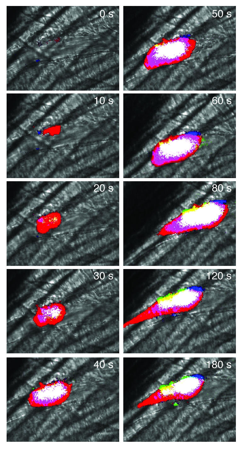
Birth of a thrombus. Intravital wide-field imaging of platelet, tissue factor, and fibrin deposition in the developing thrombus of a living WT mouse following endothelial injury. Blood flow is from right to left. Platelets, tissue factor, and fibrin were labeled using fluorescently tagged antibodies directed at CD41, tissue factor, and human fibrin, respectively. These components were imaged in 3 separate fluorescence channels. A black and white brightfield image indicates the histologic context of the composite image. To simplify analysis of the composite image, the dynamic range of the intensity of each pseudocolor was minimized. Red, platelets; green, tissue factor; blue, fibrin; yellow, platelets plus tissue factor; turquoise, tissue factor plus fibrin; magenta, platelets plus fibrin; white, platelets plus fibrin plus tissue factor.