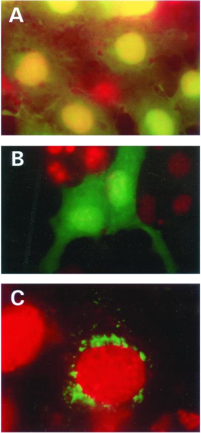
Figure 3.
Cellular localization of SIRT3 full-length and truncated proteins in Cos7 cells. (A) Shown are cells transfected with vector plasmid. Strong expression of the EGFP in the nucleus and also expression in the cytoplasm is evident. Nuclei were stained with DAPI and colored red, hence the yellow coloration when the green and red signals are superimposed in the nucleus. (B) The N-terminal (1–142 aa) deleted SIR2T3 gene fused in-frame to the EGFP reading frame was transfected into these cells. Note the diffused expression of the chimeric EGFP protein throughout the cell. Unlike with the EGFP shown above, there was no preferential expression of the chimeric protein in the nucleus. Nuclei were detected by counterstaining as described for A. (C) SIR2T3 fused to EGFP (green), localized predominantly around the nuclear periphery. Red shows the nucleus detected by counterstaining with DAPI .

