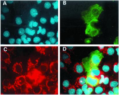
Figure 5.
Subcellular localization of truncated SIRT3 in Cos7 cells. (A) DAPI staining of cell nuclei (blue). (B) SIRT3–EGFP fusion construct transfected into the cells. Green shows the localization of the protein predominantly throughout the cytoplasm. (C) Cells stained with the mitochondrial marker, Mitotracker (red). (D) Superimposition of A–C. Note that the fusion construct does not colocalize with the mitochondrial marker.

