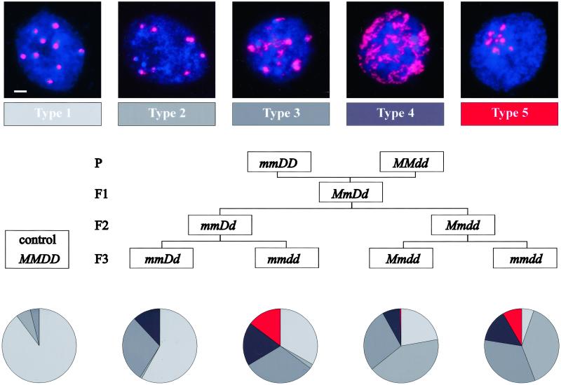
Figure 3.
Chromatin organization in interphase nuclei of single and double mutants. (Top) Classification based on distribution of the pericentromeric heterochromatin (red, 180-bp repeats revealed by fluorescence in situ hybridization analysis; blue, DNA stained with 4′,6-diamidino-2-phenylindole). (Scale bar, 2 μm.) (Middle) Genetic pedigree of the plant material. (Bottom) Representation of the five types of nuclei in different F3 genotypes. Results from 6–12 independent experiments are combined. Number of nuclei evaluated: MMDD, 829; mmDD, 1,002; mmdd, 2,257; Mmdd, 778; mmdd, 2,280.

