
Figure 3.
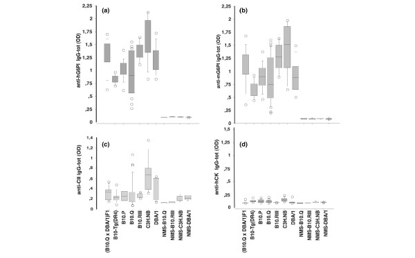
Antibody analysis. Indicated mouse strains were immunized with 200 μg human glucose-6-phosphate isomerase (hG6PI) in complete Freund's adjuvant and bled at day 40 for antibody analysis. ELISA plates were coated with (a) 10 μg/ml hG6PI, (b) mouse G6PI (mG6PI), (c) collagen type II (CII), or (d) human creatine kinase (hCK). Sera from nonimmunized mice (n = 5) of different genetic backgrounds were used as negative controls. The figures show the optical density (OD) value for total IgG responses at a serum dilution of 1:1,000 for hG6PI, mG6PI and hCK (panels a, b and d) and 1:100 for CII (panel c). The results are represented as box plots, indicating the median, the 25th and 75th centiles as boxes, and the 10th and 90th centiles as whiskers. Outliers are indicated as circles.