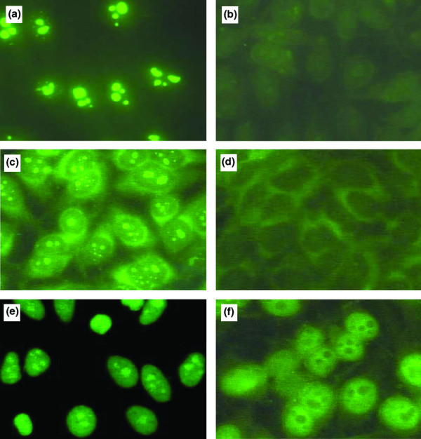
Figure 2.

Immunofluorescence staining pattern of anti-nucleophosmin-positive WB mouse and SLE sera on HEp-2 cells. An anti-nucleophosmin (anti-NPM) mouse mAb and anti-NPM antibody-positive mouse sera gave a nucleolar pattern (a,c). Anti-NPM-antibody-positive systemic lupus erythematosus (SLE) sera yielded homogeneous nuclear and nucleolar staining (e). The nucleolar staining was abrogated when the mAb and sera from mouse (b,d) or patient (f) were previously incubated with 10 μg of recombinant human nucleophosmin.
