
Figure 3.
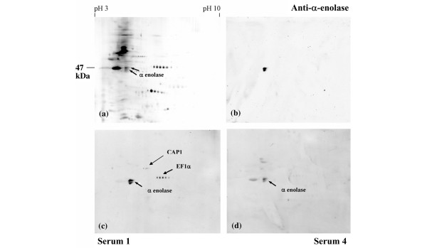
Characterisation of the 47 kDa protein by two-dimensional electrophoresis. Proteins in the 47 kDa rich monocytic S100 fraction were separated by two-dimensional electrophoresis according to charge (x-axis) and molecular mass (y-axis). (a) The full complement of proteins was observed by silver staining. (c,d) Proteins reacting with rheumatoid arthritis serum samples 1 (c) and 4 (d) were highlighted by immunoblotting. (b) The highly reactive 47 kDa protein was confirmed as α-enolase by immunoblotting with the goat anti-α-enolase antibody. CAP1, adenyl cyclase-associated protein 1; EF1α, elongation factor 1α.