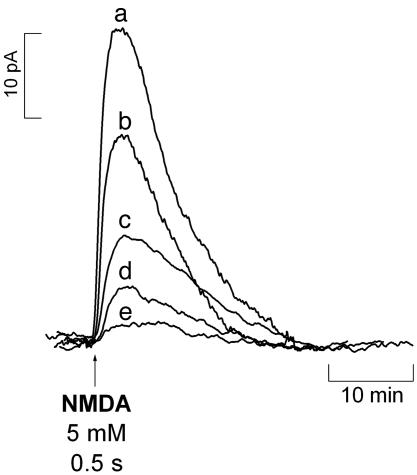
Fig. 1.
Diffusional spread of  produced upon NMDA receptor activation. The
  microssensor was inserted in the CA1 pyramidal cell layer, and the stimulation pipette was placed at increasing distances: 0 μm (a), 100 μm (b), 200 μm (c), 300 μm (d), and 400 μm (e). The stimulus consisted of a 500-ms ejection of NMDA (5 mM).

