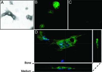
Fig. 2.
TRPV5 is predominantly visualized in the ruffled border membrane of murine osteoclasts. Murine osteoclasts derived from bone marrow were cultured on bone slices in the presence of M-CSF and RANKL for 6 days. (A) Osteoclasts were identified by TRACP staining. (B and C) TRPV5 staining (green) was demonstrated in osteoclasts derived from TRPV5+/+ mice (B) but not from TRPV5-/- mice (C). (D) Confocal laser scanning microscopy showed TRPV5 staining predominantly at the ruffled border membrane, where bone resorption occurs. Nuclei are stained with DAPI (blue).

