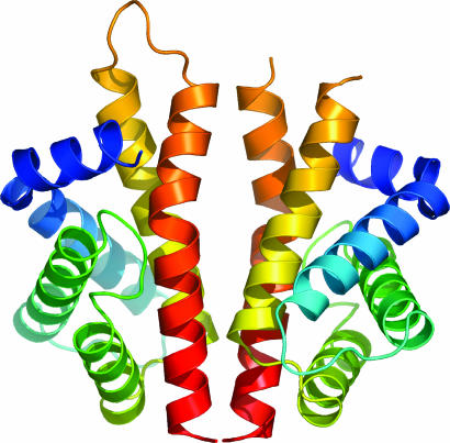
Fig. 1.
N-RsbR dimer. A protein cartoon of the structure of the dimer of N-RsbR, with each protomer in the dimer colored in a continuous color rainbow, from blue at the N terminus to red at the C terminus. The loop between residues 102 and 107 can be traced in only one of the six independent molecules in the crystallographic asymmetric unit, drawn here in orange toward the top of the left molecule only. This figure and subsequent molecular representations were prepared with pymol (http://pymol.sourceforge.net).

