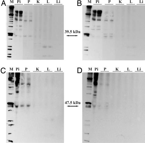
Fig. 2.
Western blot analysis of SV1-SV2-SV4 (A and B) and pGHRH-R (C and D) in samples of normal human prostate (P), kidney (K), lung (L), liver (Li), and pituitary (Pi) tissue. (A and C) Blots incubated with the purified antisera for SV1-SV2-SV4 (A) and pGHRH-R (C). (B and D) Blots with the antisera that were depleted by incubation with specific blocking peptides.

