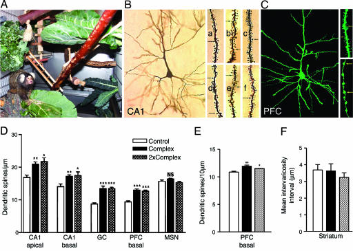
Fig. 1.
Environmental complexity enhances dendritic spine density in the adult marmoset brain. (A) Photograph of a marmoset in a complex environment, representing ≈40% of a complex single cage, with branches, vegetation, and objects typically included in the complex environment: a straw nest, a tree stump with holes, wooden swings, a wooden ladder, and blocks. (B) Photomicrograph of a Golgi impregnated CA1 pyramidal neuron, with close-up views of representative CA1 apical (a-c) and basal (d-f) dendrites, from animals in control (a and d), and complex single (b and e) and complex double cages (c and f). Arrows point to spines. (C) Photomicrograph of a DiI-labeled PFC pyramidal neuron (green color assigned for illustration purposes), with close-up views of a representative basal dendritic segment (Upper Right) and a cortico-striatal axonal segment (Bottom Right). Arrows point to spines and varicosities, respectively. (D) Marmosets living in complex environments for 4 weeks have greater dendritic spine density on several types of Golgi-impregnated neurons in the hippocampus and the PFC, compared with marmosets living in standard laboratory environments. Error bars represent SEM; asterisks reflect statistically significant differences from control group on Tukey post hoc comparison: *, P < 0.05; **, P < 0.01; ***, P < 0.001. (E) Marmosets living in complex environments have greater dendritic spine density on DiI-labeled neurons compared with animals living in standard laboratory conditions. (F) No differences in intervaricosity spacing on cortico-striatal axons were observed for marmosets living in standard and two types of complex housing.

