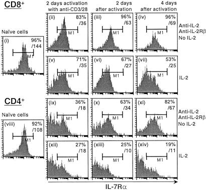
Figure 6.
IL-7Rα expression in the presence or absence of IL-2 after anti-CD3/CD28 activation. Naive CD8+ or CD4+ splenocytes (i and viii) were isolated from wild-type mice and activated in anti-CD3-bound plates in the presence of anti-CD28-, anti-IL-2-, and anti-IL-2Rβ-neutralizing antibodies (both at 20 μg/ml; ii and ix, with antibodies and no IL-2) or in the presence of anti-CD28 and IL-2 for 2 days (v and xii, with IL-2). Cells then were washed, and the neutralizing antibodies were refreshed. The cells were cultured for another 2 (iii, vi, x, and xiii) or 4 days (iv, vii, xi, and xiv). Cell-surface IL-7Rα expression was determined by flow cytometry. The left boundary of the M1 region was set based on isotype-matched control antibody. Values in the upper right corners are the percentages of cells expressing high levels of IL-7Rα (M1 gate) and overall fluorescence intensity of cell-surface IL-7Rα.

