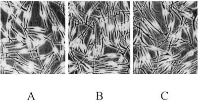
Fig 1.
Melanocytes derived from skin of a control (A), from normal skin of a NF1 patient (B), and from a NF1 café-au-lait macule (C). (×75.) Melanocytes of five controls and NF1 patients (skin and café-au-lait macule) were cultured in parallel. At day 7, 20 randomly taken pictures were digitized (450–600 cells per culture). The different morphology of NF1 cells compared with the normal melanocytes is obvious.

