
Figure 2.
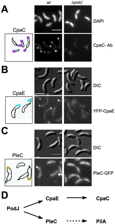
PodJ is required for the polar localization of the CpaC secretin, the CpaE pilus assembly factor, and the PleC histidine kinase. The location of each protein was examined in wild-type (wt) and ΔpodJ mutant cells. (A) Indirect IFM using affinity-purified CpaC antibody (Ab). Shown are images of 4′,6-diamidino-2-phenylindole (DAPI)-stained chromosomal DNA (Upper), IFM images of CpaC (Lower), and a drawing with the location of CpaC (purple dots) in wild-type cells. (B) CpaE location observed in cells expressing YFP–CpaE. Shown are differential interference contrast (DIC) images (Upper), fluorescence images (Lower), and a drawing with the location of YFP–CpaE (blue dots) in the podJ+ yfp-cpaE strain. (C) PleC location observed in cells expressing PleC–GFP. Shown are DIC images (Upper), fluorescence images (Lower), and a drawing with the location of PleC-GFP (gold dots) in the podJ+ pleC-gfp strain. (D) The parallel pathways of PodJ localized proteins. (Scale bars, 2 μm.)