Figure 3.
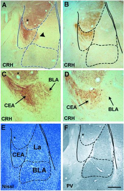
CRH is released from CEA neurons after footshock administration in an inhibitory avoidance task. Extracellular immunoreactive CRH in the CEA of rats killed 30 min after receiving the higher-intensity footshock (0.60 mA, 1.5 s) is enhanced (A and C) compared with stress-free controls (B and D) and appears to invade the BLA. Boundaries of the BLA are delineated using Nissl stain (E). Further illustration of the parvalbumin (PV)-free CEA is provided (F); note that only a portion of the lateral nucleus (La) expresses PV. Overlay is according to Paxinos and Watson (34). [Bar = 350 μm (A, B, E, and F) and 150 μm (C and D).]

