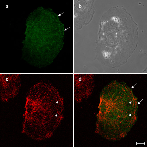
Figure 5.

Overexpressed aurora B is not colocalized with phosphorylated myosin II regulatory light chain in interphase NRK cells. An NRK cell overexpressing aurora B-GFP was stained with antibodies that specifically recognised myosin II regulatory light chain phosphorylated at Ser19 and then examined the subcelluar localization of aurora B-GFP (a) and phopshorylated myosin II regulatory light chain (c) by confocal laser microscopy. Corresponding phase and merged images (green; aurora B-GFP, red; phosphorylated myosin II regulatory light chain) are shown in panels b and d, respectively. Overexpressed aurora B is associated with the cortex (a and d, arrows), while phosphorylated myosin II regulatory light chain is enriched in the cell periphery (c, and d, arrowheads). Bar, 10 μm.
