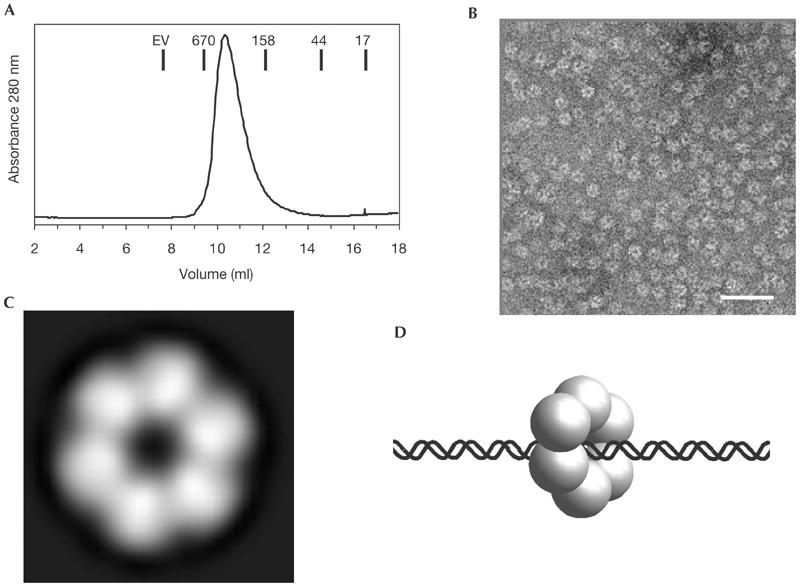
Figure 5.

Structure of MlaA. (A) Gel filtration chromatogram showing that MlaA has a hydrodynamic radius corresponding to approximately 360-kDa molecular weight. Molecular weight standards are indicated. EV: exclusion volume. (B) Negative stain electron micrographs of MlaA show multimeric ring-shaped particles (bar: 50 nm). (C) Averaging over 1,560 electron micrograph particles shows that MlaA forms approximately 120 Å large hexameric rings with a central hole of approximately 20–30 Å diameter. This structure resembles AAA ATPases and further indicates that MlaA could be a DNA acting ATPase in DNA recombination. (D) Model for an MlaA:DNA complex with DNA threaded through the central hole.
