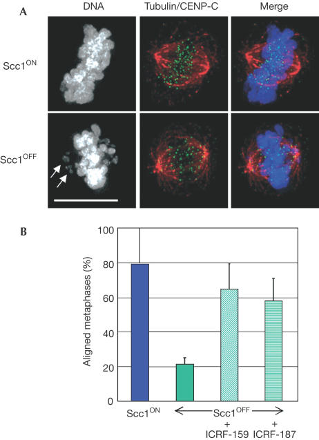
Figure 2.
Successful alignment of Scc1-deficient chromosomes at a metaphase plate following treatment with topo II inhibitors. (A) A metaphase cell was defined as a cell with its chromosomes all aligned on the plate, as shown at the top, whereas cells with chromosomes scattered about the plate (arrows, lower panels) were defined as prometaphase. Cells on polylysine-treated slides were fixed and incubated with antibodies to α-tubulin (red) and CENP-C (green), along with DAPI (grey in left panels). Scale bar, 10 μm. (B) Histogram showing the relative percentage of metaphase Scc1OFF cells following the treatment of synchronized cells with the topo II inhibitors ICRF-159 or ICRF-187 for 1 h before analysis. Cells in which the spindle axis was not parallel to the slide were excluded from this analysis. Data shown are the mean±standard deviation of at least three experiments in which at least 25 cells with appropriately aligned spindles were scored blind. Levels of alignment under all conditions differed significantly from those observed in Scc1OFF conditions (χ2-test; P<0.01). A control experiment on 25 Scc1ON metaphases showed 85 and 84% aligned at metaphase after treatment with ICRF-159 and ICRF-187, respectively.

