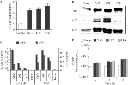
Figure 3.
Signal transduction by CpG-ODN, LPS and LTA. (A) RAW264.7 cells were transiently transfected with an NFκB-dependent reporter plasmid. Cells were stimulated with 100 nM CpG-ODN, 100 ng/ml LPS or 30 μg/ml LTA for 7 h, and luciferase induction was determined. (B) RAW264.7 cells were stimulated as above for 15 min. Similar amounts of cellular extracts were blotted with phosphospecific antibodies for p38, JNK or ERK MAP kinase. (C) Peritoneal macrophages from IRF1−/− and IRF1+/− mice were stimulated as above. Supernatants were assayed for TNFα and IL-12p40. (D) RAW264.7 macrophages were stimulated as above for the indicated time periods. ICSBP mRNA expression was measured by quantitative RT-PCR.

