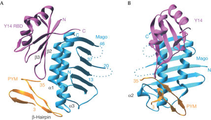
Figure 2.
Structure of the Drosophila PYM–Mago–Y14 ternary complex. (A) View of the complex between the RBD domain of Y14 (pink), Mago (cyan) and the N-terminal domain of PYM (orange). PYM binds at the edge of the Y14 β-sheet (β2–β3 loop) and at the edge of the Mago α-helices, spanning both components of the heterodimer. Dotted lines represent the approximate path of loops in Mago that were disordered and not modelled. (B) View of the complex after a 90° rotation about the vertical axis with respect to the view in (A).

