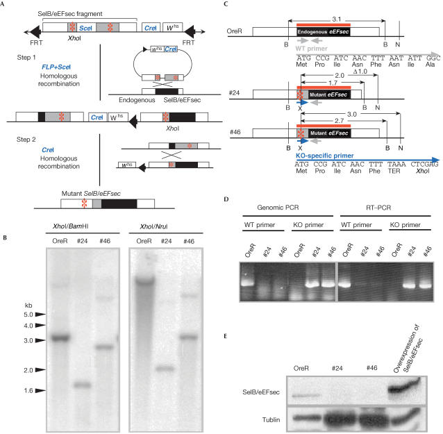
Figure 2.
Schematic representation of SelB/eEFsec mutagenesis by homologous recombination. (A) The targeting DNA fragment excised by FLP from the donor construct is expected to recombine with endogenous SelB/eEFsec locus (step 1; for details, see Rong & Golic, 2000; Rong et al, 2002). In a second step, the tandem duplication was reduced to a single copy by a homologous recombination event due to a CreI-mediated double-strand break (step 2). Red asterisks: introduced TAA stop codon. (B) Southern blot with genomic DNA from wild-type and homozygous knockout mutant (KO#24 and KO#46) individuals. DNA was digested by XhoI, which is the newly generated site only in mutant DNA (see (A)), and by either BamHI or NruI. The SelB/eEFsec-specific DNA hybridization probe is indicated by red lines. (C) Gene structure of the wild-type (shown on top) and of SelB/eEFsec mutant alleles (#24 and #46). Restriction sites are as follows: B (BamHI), N (NruI) and X (XhoI). Size of DNA fragments in kilobase pairs (kb). (D) Genomic DNA and total RNA were prepared from wild-type (OreR) or homozygous knockout mutant flies (KO#24 and KO#46). Wild-type and knockout-specific primers are shown in (C); for their sequence, see Material and methods. (E) Anti-SelB/eEFsec antibody staining of western blots loaded with protein extracts of wild-type flies (OreR), homozygous knockout mutant flies (KO#24 and KO#46) or flies overexpressing SelB/eEFsec from a cDNA-derived transgene (see text). Note that SelB/eEFsec is left undetected in the KO#24 and KO#46 mutant individuals even in the presence of higher amounts of loaded protein (see loading control provided by anti-α-tublin staining).

