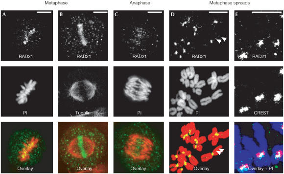
Figure 1.
Immunostaining of RAD21/SCC1 antibodies in HeLa cells. Images were obtained using RAD21 Ab2. Chromosomes were counterstained using propidium iodide (PI). (A) Metaphase showing RAD21/SCC1 (green) and chromosomes (red), (B) co-labelling of RAD21/SCC1 (green) with tubulin (red), (C) anaphase, (D) metaphase chromosome spreads (note the residual RAD21/SCC1 staining between sister chromatids) (arrowheads) and (E) co-labelling of RAD21/SCC1 (green) with CREST-6 sera for centromeres (red) on metaphase chromosome spreads (purple). Scale bar, 10 μm.

