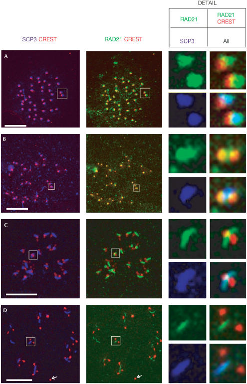
Figure 4.
RAD21/SCC1 persists at the centromeres of meiotic chromosomes until anaphase II. Metaphase I to anaphase II chromosome spreads of spermatocytes were immunostained with antibodies against SCP3 (purple), CREST (staining for kinetochores, red) and RAD21 (green). (A) Metaphase I, (B) anaphase I, (C) metaphase II and (D) metaphase II/anaphase II; the arrow indicates the loss of SCP3 and RAD21/SCC1 staining from centromeres. Detailed views of chromosomes (boxed, not to scale) are RAD21/SCC1 alone (top left panel), RAD21/SCC1 and CREST merged (top right panel), SCP3 alone (bottom left panel) and all three proteins merged (bottom right panel). Colocalization of RAD21 with CREST is yellow in a merged image. Light blue indicates colocalization of all three proteins. Scale bar, 10 μm.

