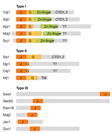
Figure 2.
Structural classification of J proteins. Representation of type I, II and III J proteins from yeast aligned according to the amino-terminus of the mature protein. Mdj1 and Scj1 have targeting sequences processed in the mitochondria and endoplasmic reticulum, respectively. The grey boxes represent each polypeptide and show the scale of the J domain (J, orange), glycine-rich region (G, yellow) and zinc-finger domain (Zn-finger, green) found in some J proteins compared with the protein–protein-interaction domains that bind non-native substrate. CTD, carboxy-terminal domain; TM, transmembrane segment.

