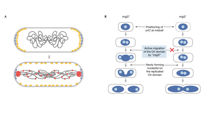
Figure 2.
Models of the B. subtilis and E. coli 'centromeres'. (A) In B. subtilis, RacA protein (red) interacts with DivIVA (blue) during sporulation to pull the origin regions to cell poles in the predivisional sporangium, as MinCD (yellow) moves away. (Figure provided by R. Losick; Ben-Yehuda et al, 2003.) (B) In E. coli, a migS+ and migS− E. coli cell model of the effect of migS on polarization of oriC. (Figure provided by H. Niki; Yamaichi & Niki, 2003.)

