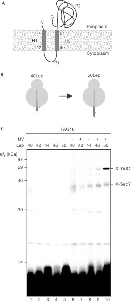
Figure 1.
Initial contacts of Lep H1 during membrane insertion. (A) Topology of Lep in the inner membrane. (B) Schematic representation of the 40 and 50Lep constructs with a crosslinking probe at position 10. The transmembrane regions are presented as thick lines. (C) In vitro translation of nascent Lep 40–50-mer was carried out in the presence of inner membrane vesicles and the (Tmd)Phe-tRNAsup. After translation, samples were UV irradiated to induce crosslinking and extracted with sodium carbonate.

