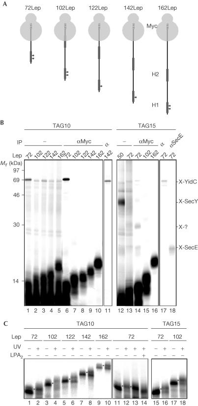
Figure 2.
Consecutive interactions of Lep H1 during membrane insertion. (A) Nascent Lep species used to study insertion of H1. The photo-crosslinking probes are indicated (positions 10 and 15). The C-terminal Myc tags are indicated as hatched bars. (B) In vitro translation, crosslinking and carbonate extraction of nascent Lep 50–162-mer were carried out as described in Fig 1. Carbonate-insoluble material was immunoprecipitated as indicated. (C) Nascent Lep 50–162-mer was produced, crosslinked or kept in the dark, immunoprecipitated using anti-Myc serum and analysed by 15% SDS–PAGE. To identify lipid crosslinking adducts (indicated by asterisks), membranes of photo-crosslinked 72LepTAG10 samples were not carbonate extracted but spun through a highsalt sucrose cushion and incubated with bee venom phospholipase A2 (PLA2) or mock treated with incubation buffer (lanes 13 and 14).

