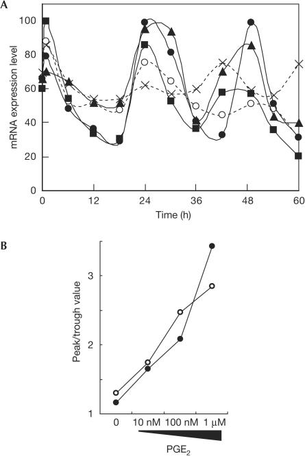
Figure 2.
Circadian gene expression induced by prostaglandin E2 (PGE2) in NIH3T3 cells. (A) Circadian oscillation of the expression level of mPer2 mRNA induced by varying concentrations of PGE2. At time 0, NIH3T3 cells were treated with no stimulus (crosses), PBS (open circles) and 10 nM (squares), 100 nM (triangles) and 1 μM (filled circles) of PGE2, and the mRNA expression levels were monitored by the real-time quantitative PCR method. Each value was normalized to mG3PDH. Data shown are representative of three independent experiments. (B) Robustness of circadian gene expression induced by PGE2 in NIH3T3 cells. The peak and trough values of mRNA expression levels in the second cycle of oscillation were read from the graph and the extent of amplitude was plotted. Graphs shown are data for mPer2 (filled circles) and mDBP (open circles).

