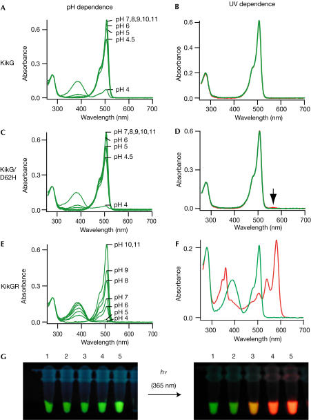
Figure 2.
Spectral properties of KikG variants. (A,C,E) pH dependence of absorption spectra of KikG (A), KikG/D62H (C) and KikGR (E). (B,D,F) Absorption spectrum at pH 7.0 acquired before (green line) and after (red line) irradiation with ∼365 nm light (1.8 mW cm−2) for KikG (B), KikG/D62H (D) and KikGR (F). (G) Fluorescence of protein solutions of KikG mutants acquired immediately after (left) and several minutes after (right) excitation with ∼365 nm light. 1, KikG; 2, KikG/D62H; 3, KikG/M40V/D62H/I198M; 4, KikG/M10I/L12V/M40V/V60A/D62H/Y119N/P144S/R197L/I198M; 5, KikGR.

