Figure 2.
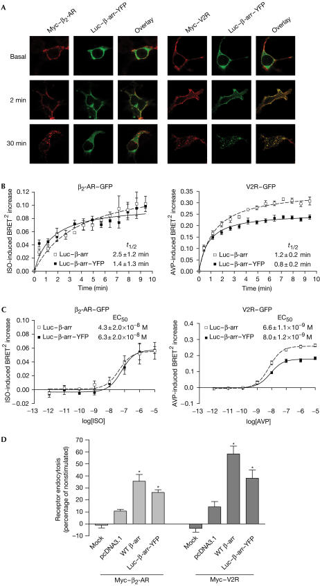
Functionality of double-brilliance β-arr. HEK293 (A–C) or COS (D) cells were transiently transfected with the indicated plasmids. (A) Cells incubated or not in the presence of saturating concentrations of specific agonists (β2-AR, 10 μM isoproterenol (ISO); V2R, 1 μM arginine vasopressin (AVP)). Localization of Luc–β-arr–YFP and Myc-tagged receptors was analysed by confocal fluorescence microscopy. (B) Agonist-induced recruitment of β-arr measured using BRET2. t1/2=half-time of maximal β-arr recruitment. (C) Dose-dependent recruitment of β-arr to the receptors measured in BRET2 following 2 min stimulation with the agonist. EC50=concentration of agonist producing half-maximal β-arr recruitment. (D) Cells treated or not for 15 min with the specific agonists at 37°C and cellsurface receptor levels measured by enzyme-linked immunosorbent assay (ELISA). Receptor endocytosis is defined as the loss of cell-surface immunoreactivity and is expressed as a percentage of total immunoreactivity measured under basal conditions. Expression levels of β-arr were controlled using western blot (data not shown). Data are the mean±s.e.m. of at least three independent experiments. *P<0.05 between treatment and each individual control condition. Mock, nontransfected cells.

