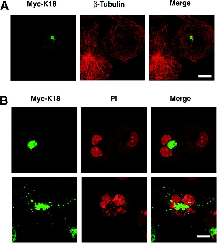
Figure 6.
Disruption of the microtubular array by K18 aggregates. (A) Impaired function of microtubules in cells containing K18 aggregates. COS-7 cells that had been transfected with a plasmid encoding Myc-K18 were stained with antibodies to Myc (green; left) and to β-tubulin (red; middle). (B) Abnormal morphology of the nucleus of cells containing K18 aggregates. COS-7 cells containing Myc-K18 (green) aggregates were also stained with PI (red) to detect nuclei. Superimposed images are shown (merge). A cell with K18 aggregates contains two nuclei (top), whereas another cell contains fragmented nuclei (bottom). Bar, 20 μm.

