Figure 4.
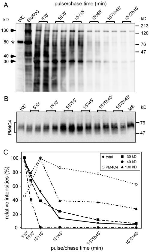
Recycling of plasma membrane components is operated in distinct steps. Before uptake of LBs, cell surface proteins were labeled with a biotinylated cross-linker. Extracts and purified phagosomes were separated by SDS-PAGE and blotted onto a membrane. Detection was carried out by avidin-HRP (A). In whole-cell extracts of nonbiotinylated cells (WC), very low background is visible, except an endogenously biotinylated 80-kDa protein (asterisk). After biotinylation, dozens of bands appear, with a particularly prominent one at 130 kDa. (B) The blot was then stripped and probed with an antibody against a plasma membrane glycoprotein (PM4C4). Signals were quantified by chemiluminescence imager, and the average of doublets was plotted as a function of the pulse/chase time (C). Total indicates that the signal was integrated over the whole lane, and 30 kDa, 40 kDa, and 130 kDa indicate three major biotinylated bands that behaved with distinct kinetics.

