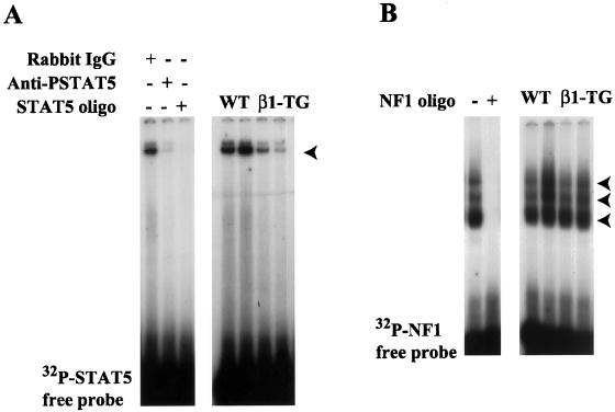
Figure 5.
Detection of STAT5 and NF1 in the nuclear extracts from wild-type and MMTV-β1-cyto mammary glands by band-shift analysis. (A) Left, 1 μg of nuclear extract from a 2-d wild-type-involuting gland was incubated with either 1 μg of rabbit IgG, 1 μg of anti-phospho-STAT5 antibody, or a 50-fold excess of unlabeled STAT5 oligonucleotide, before adding the 32P-labeled STAT5 probe. Right, 1 μg of nuclear extracts from two wild-type and two MMTV-β1-cyto transgenic glands was incubated with the 32P-labeled STAT5 probe. The arrowhead indicates the STAT5-DNA complex. (B) Left, 1 μg of nuclear extract from a 2-d wild-type-involuting gland was incubated with the 32P-labeled NF1 probe either in the presence (+) or absence (−) of a 50-fold excess of unlabeled NF1 oligonucleotide. Right, 1 μg of nuclear extracts from two wild-type and two MMTV-β1-cyto transgenic glands was incubated with the 32P-labeled NF1 probe. As described previously, three major NF1-DNA complexes indicated by arrowheads were detected in the mammary gland nuclear extracts (Furlong et al., 1996).

