Figure 10.
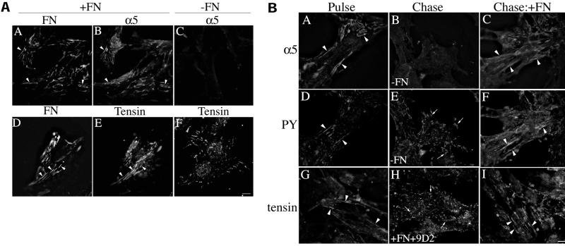
(A) α5β1 and tensin localize to fibrillar adhesions only in the presence of fibronectin. Fibronectin-null cells were incubated in the absence (C and F) or presence of 20 nM FITC-fibronectin (D and E) or 20 nM unlabeled fibronectin (A and B). After an overnight incubation, cells were fixed and permeabilized and then incubated with antibodies to fibronectin and α5 integrin (A–C) or antibodies to tensin (D–F). Fibronectin staining is shown in A and D; α5 integrin staining in B and C; and tensin staining in E and F. The same fields of view are shown in A and B and in D and E. Areas of colocalization of fibronectin and α5 integrin, or fibronectin and tensin are shown by the arrowheads. Bar, 10 μm. (B) Fibronectin matrix stability regulates maintenance of cell–matrix fibrillar adhesions. Fibronectin-null cells were incubated with 20 nM fibronectin. After an overnight incubation, cells were washed and then incubated in the absence (Chase; B and E) or presence of fibronectin (C, F, H, and I) for 23 h. In H, cells were coincubated with fibronectin and 9D2 IgG. Cells were fixed, permeabilized, and then coincubated with antibodies to α5 integrin (A–C) and phosphotyrosine (D–F) or antibodies to tensin (G–I). Panels A and D, B and E, and C and F are the same fields of view. Fibrillar adhesions are shown by the arrowheads and focal contacts by the arrows. Bar, 10 μm.

