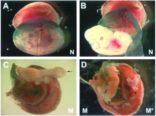
Figure 5.
Tetraploid aggregations. (A and B) Photographs of an E12.5 normal littermate control (N) embryo with whole-mount X-gal staining (blue) marking the wild-type tetraploid cells contributing to the visceral endoderm of the yolk sac. There are no tetraploid cells seen contributing to the embryo proper (B). (C) Photographs of an E12.5 mutant (M, Snx1-/-;Snx2-/-) embryo with whole-mount X-gal staining marking the wild-type tetraploid cells contributing to the visceral endoderm of the yolk sac. Note that this embryo has already arrested. An abnormal tail region is seen (arrow). (D) On the left is the mutant embryo (M, Snx1-/-;Snx2-/-, arrow) seen in C (a portion of the abnormal tail has been removed for genotyping). On the right is a mutant embryo (M*, Snx1-/-;Snx2-/-) obtained by natural matings. This embryo is representative of the most well-developed stage mutants typically achieve (arrowhead).

