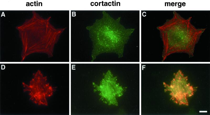
Figure 1.
Tenascin-C induces a distinct cytoskeletal organization. NIH 3T3 fibroblasts were allowed to interact with fibrin-FN (A–C) or fibrin-FN+tenascin-C (D–F) matrices for 1 h before fluorescent staining for actin with rhodamine-labeled phalloidin (A and D) and for cortactin with an anticortactin mAb (B and E). Sites of colocalization are shown in yellow (C and F). Bar, 10 μm.

