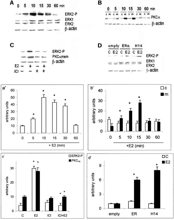
Figure 4.
MAP kinase and PKC-α pathways activation in HepG2 and HeLa cells. Time course of ERK-2 phosphorylation (a) and PKC-α translocation to the membranes (b) in HepG2 cells were performed by Western blot analysis, as described in MATERIALS AND METHODS, on control (0) and on estradiol-treated cells (E2) (10 nM) at different times. The amounts of protein were normalized by comparison with ERK-1 and ERK-2 or β-actin level expression (a′ and b′, densitometric analysis). Western blot analysis both of ERK-2 phosphorylation and PKC-α levels on membranes after pretreating 15 min with ICI 182,870 (1 μM) (ICI) on control (−) and on 10-min estradiol-treated HepG2 cells (E2) (10 nM) (c) and of ERK-2 phosphorylation on control (C) and on 10-min estradiol-treated HeLa cells (E2) (10 nM) (d) were performed as described in MATERIALS AND METHODS. The amounts of protein were normalized by comparison with ERK-1 and ERK-2 or β-actin level expression (c′ and d′, densitometric analysis). Data are the means ± SD of three independent experiments. *P < 0.001, compared with respective control values (C), determined using Student's t test. °P < 0.001, compared with respective E2 values, determined using Student's t test.

