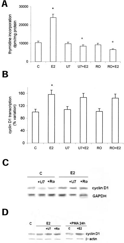
Figure 6.
Role of PKC-α in cyclin D1 gene transcription and DNA synthesis in HepG2 cells. Tymidine incorporation into DNA has been evaluated as described in MATERIALS AND METHODS on HepG2 cells pretreated 15 min with the PKC inhibitor Ro31-8220 (Ro) (1 μM) and then stimulated 6 h with vehicle (C) or E2 (10 nM) (a). Luciferase assay detection on HepG2 cells transfected with pXP2-D1-2966-luciferase and then stimulated 6 h with vehicle (C) or E2 (10 nM), after 15 min of pretreatment with the PKC inhibitor Ro31-8220 (Ro) (1 μM) or PLC inhibitor U73122 (U7) (1 μM) (b). Data are expressed as disintegrations per minute per total protein extracted and as percentage variation with respect to the controls and are the means ± SD of four independent experiments. *P < 0.001, compared with respective control values (C), determined using Student's t test. °P < 0.001, compared with respective E2 values, determined using Student's t test. Northern (c) and Western (d) blot analysis of cyclin D1 were performed as described in MATERIALS AND METHODS on control (C) and on E2-stimulated HepG2 cells (10 nM) pretreated with the PKC inhibitor Ro31-8220 (Ro) (1 μM) or PLC inhibitor U73122 (U7) (1 μM) and PMA (10 μM), respectively. The amounts of mRNA and protein levels were normalized by comparison with GAPDH and β-actin expression. Shown are typical blots of three independent experiments.

