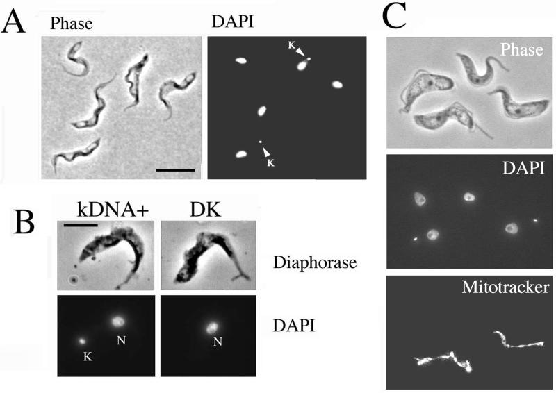
Figure 6.
Dyskinetoplastid cells can develop to stumpy forms. (A) Phase-contrast/DAPI image of parasites 16 h after acriflavine dosage of the host. Cells with (arrowed) and without a kinetoplast are shown. Both normal (kDNA+) and DK cells show stumpy morphology. Bar, 15 μm. (B) Diaphorase staining of a kDNA+ (left) and a dyskinetoplastid cell (right). Both cells are morphologically stumpy and demonstrate clear diaphorase staining along the mitochondrion, a cytochemical characteristic of stumpy forms. Bar, 10 μm. (C) kDNA+ and dyskinetoplastid cells are shown, all with stumpy morphology. Only the kDNA+ cells exhibit detectable mitochondrial staining with Mitotracker Red CMX ROS.

