
Figure 1.
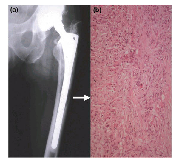
Radiographic and histologic findings in periprosthetic osteolysis and loosening of the prosthesis. (a) The radiograph demonstrates periprosthetic bone erosions along both the medial and lateral endosteal bone surfaces. The femoral head is eccentrically placed in a superior position in the acetabular cup, indicating polyethylene wear and the generation of particles. (b) The bone in the osteolytic lesions is replaced by fibro-inflammatory tissue (arrow) consisting of a background of fibroblasts with a diffuse infiltrate of inflammatory cells (lymphocytes, plasma cells, and macrophages), which is most intense in the top left-hand quadrant of this micrograph. Released particles of wear debris accumulate in this tissue, which acts as a reservoir for them and thus enhances the progression of the bone loss and further loosening. This patient underwent a revision arthroplasty.