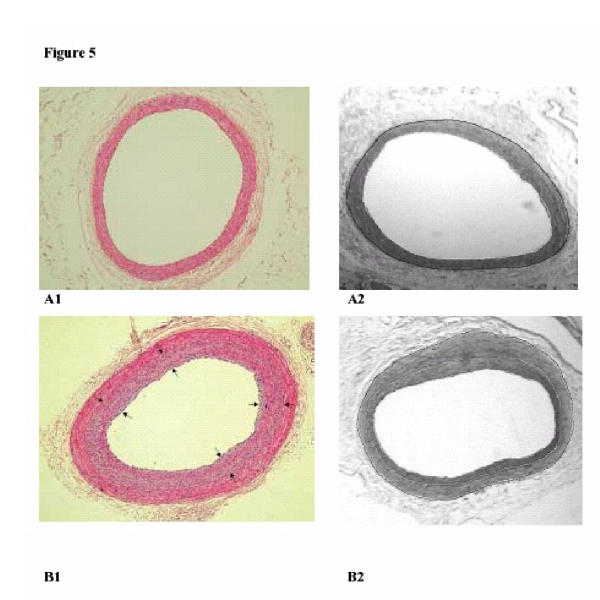
Figure 5.

Photographs in the left column show HE-stained cross-sections of the iliac artery at 0 weeks (A1) and at 16 weeks (B1) after balloon injury (BI). The area between the black arrows indicate the area intimal hyperplasia. The digital planigraphs in the right column show the iliac artery at 0 weeks (A2) and at 16 weeks (B2) after BI. The white lines demarcate the IH area and the black line represents the external elastic lamina (border medial area = area between black and first white line).
