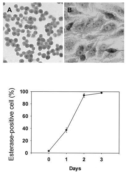
FIG. 3.
Nonspecific esterase staining of IFN-γ-induced macrophage-like cells. TH2.52 cells were cultured with IFN-γ (100 U/ml), and the time course in the appearance of esterase-positive cells was monitored for 4 days (top). A typical staining pattern of esterase-positive cells at day 4 is shown (bottom). (A) Untreated control cells; (B) IFN-γ-treated cells. Magnification, ×140.

