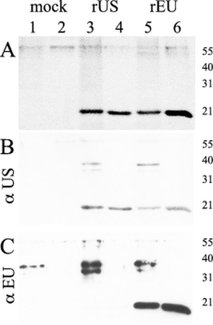
FIG. 3.
Analysis of affinity-purified recombinant N-proteins by SDS-PAGE and Western blot analysis. (A) Coomassie blue staining of an SDS-15% polyacrylamide gel. Crude cell lysates (lanes 1, 3, and 5) and affinity-purified proteins (lanes 2, 4, and 6) for mock antigen (lanes 1, 2), rUS-N-protein (lanes 3, 4), and rEU-N-protein (lanes 5, 6) are shown. (B and C) Western blot analysis was performed for the same samples with either US PRRSV-specific antiserum US364/d42 (B) or EU PRRSV-specific antiserum EU8787/d42 (C). The sizes of marker proteins in kilodaltons are indicated.

