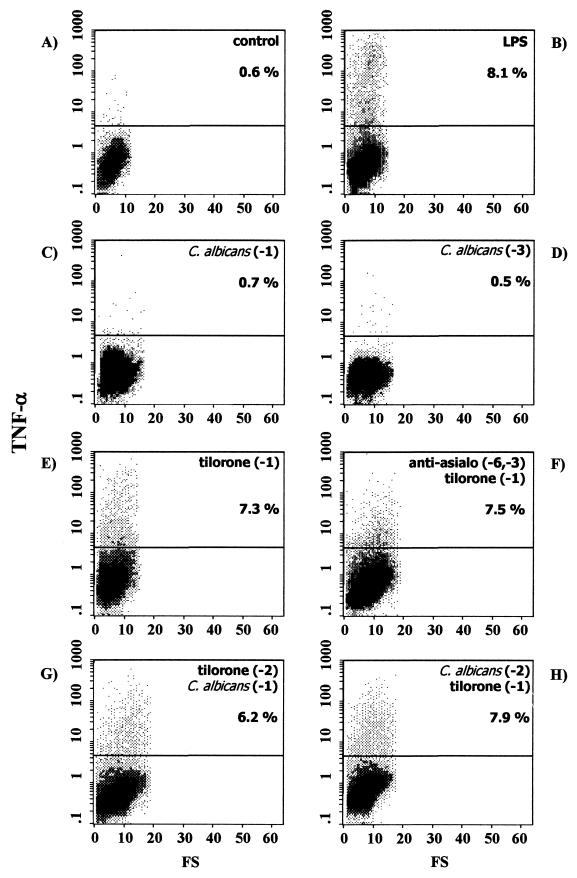
FIG. 2.
Intracellular TNF-α expression in splenic macrophages from control naïve BALB/c mice (A) or BALB/c mice exposed to different stimuli, as follows: in vitro LPS stimulation (B), C. albicans infection 24 h before the assay (C), C. albicans infection 72 h before the assay (D), tilorone treatment 24 h before the assay (E), depletion of NK cells by intraperitoneal injection of anti-asialo-GM1 on the indicated days, followed by tilorone treatment 24 h before the assay (F), tilorone treatment 48 h before the assay, followed by i.v. challenge with live C. albicans 24 h before the assay (G), and i.v. C. albicans infection 48 h before the assay, followed by tilorone treatment 24 h before the assay (H). Percentages reflect cytokine-positive cells. Results are from one representative experiment out of three performed with cells from different mice.

