
FIGURE 1.
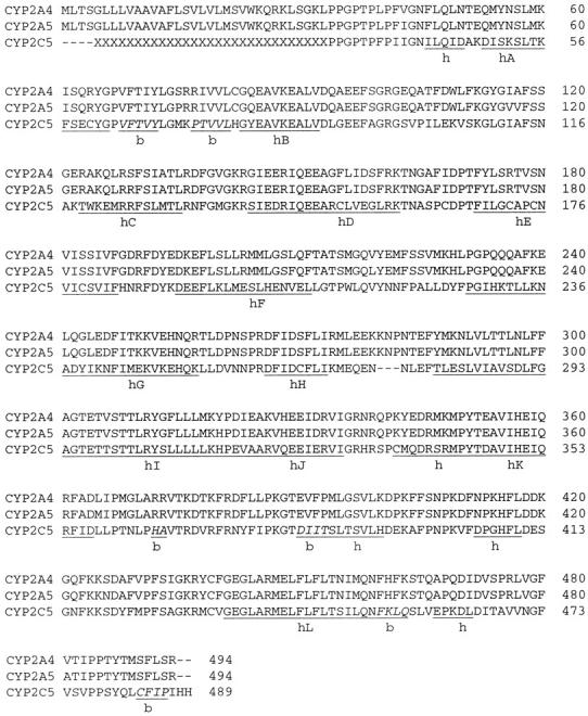
Sequence alignment of CYP2A4, CYP2A5, and CYP2C5. Residues experimentally shown to modulate substrate specificity (Ala-117, Leu-209, Leu-365, and Val-481 in CYP2A4) are shown in gray (Negishi et al., 1996). Thr-305, conserved in bacterial and mammalian CYP450s, is also highlighted. The alignment was performed using Clustal X (Thompson et al., 1997). Helices and β-sheets are labeled with letters h and b, respectively. Additionally, helices A-L are identified, and residues comprising β-sheets are labeled in italics (as assigned by Williams et al., 2000a).