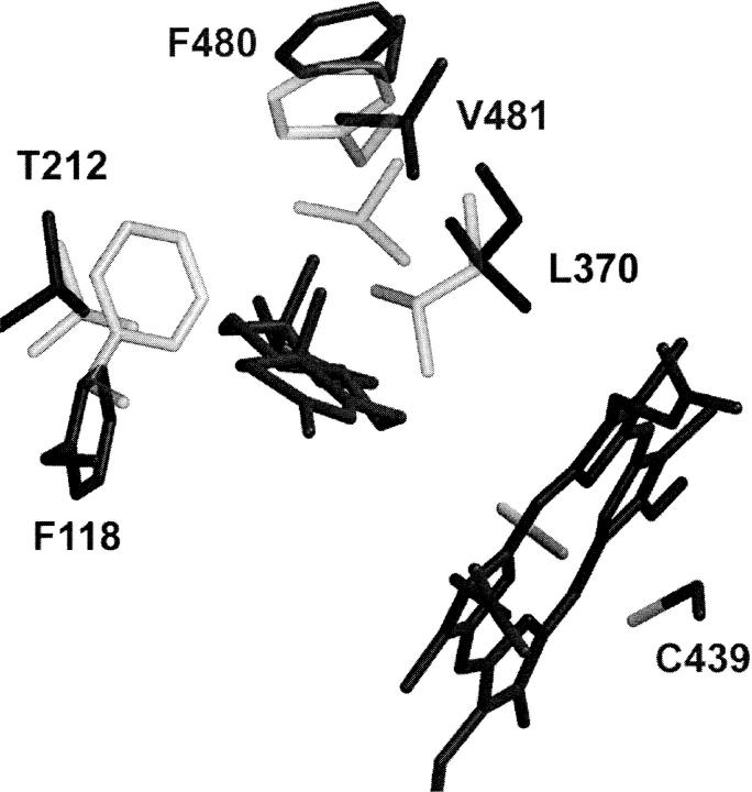
FIGURE 5.
The active site of solvent equilibrated CYP2A4 (light) and CYP2A4/T (dark). Only the residues lining the active site (within 4 Å of the docked testosterone in CYP2A4/T) that showed significant change are shown. The hemes of the two structures were overlaid; only the CYP2A4/T heme is shown.

