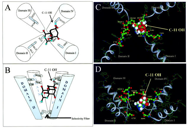
FIGURE 5.
(A and B) Schematic emphasizing the orientation of TTX in the outer vestibule as viewed from top and side, respectively. The molecule is tilted with the guanidinium group pointing toward the selectivity filter and C-11 OH forming a hydrogen bond with D1532 of domain IV. (C and D) TTX docked in the outer vestibule model proposed by Lipkind and Fozzard (Lipkind and Fozzard, 2000). The docking arrangement is consistent with outer vestibule dimensions and explains several lines of experimental data. The ribbons indicate the P-loop backbone. Channel amino acids tested are in ball and stick format. Carbon (shown as green); nitrogen (blue); sulfur (yellow); oxygen (red); and hydrogen (white).

