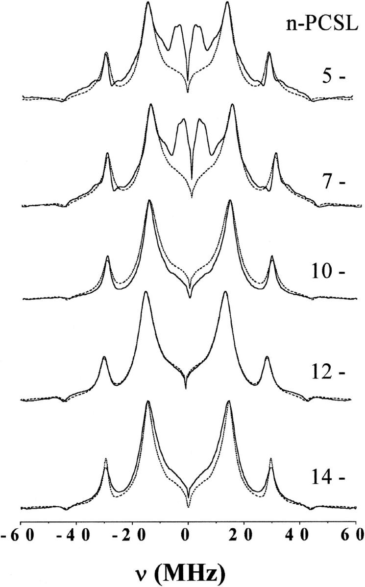
FIGURE 3.

Fourier transform of the relaxation-corrected echo decays from n-PCSL spin-labeled phosphatidylcholines in bilayer membranes of dipalmitoyl phosphatidylcholine with 50 mol% cholesterol. Solid lines are for membranes dispersed in D2O and dashed lines are for membranes dispersed in H2O. The position, n, of spin labeling in the sn-2 chain is indicated on the figure. Spectra are normalized to the maximum peak height.
