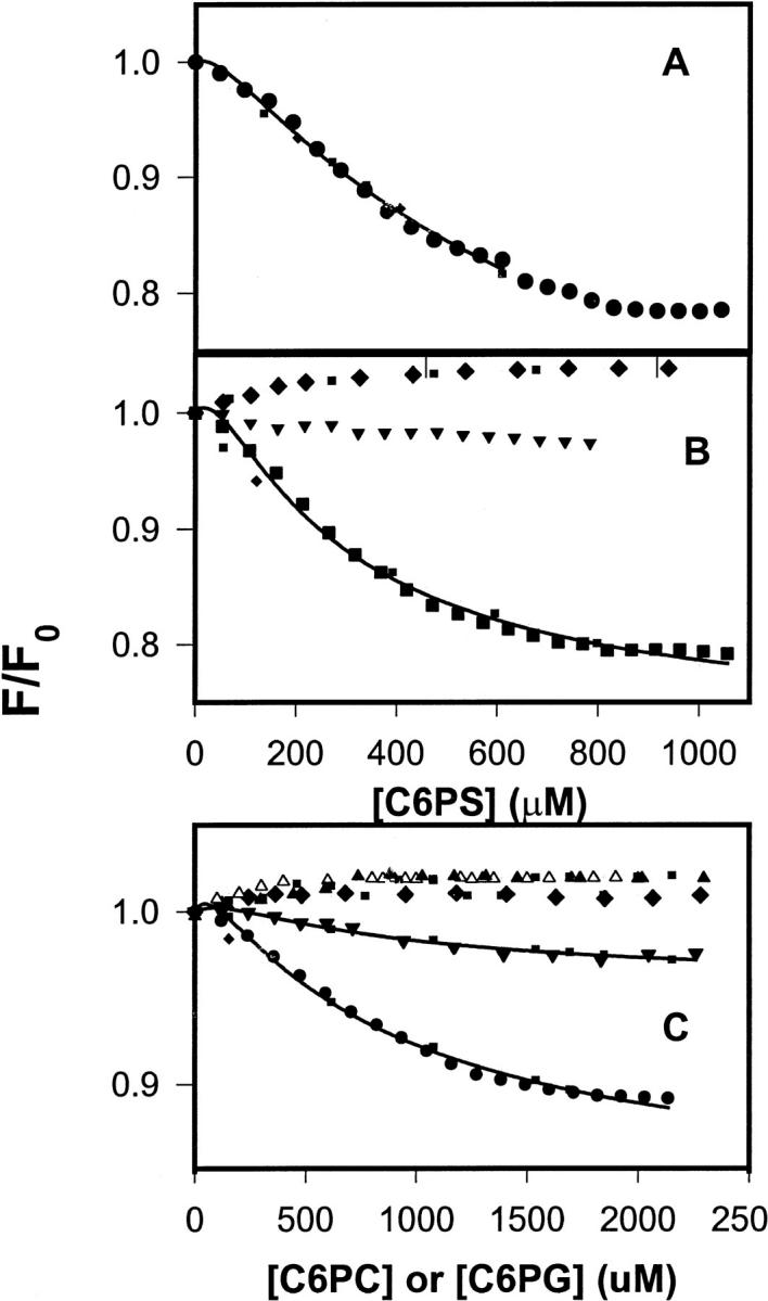
FIGURE 1.

(A) DEGR-Xa (filled circles) at 100 nM in buffer A (50 mM Tris, 0.1 M NaCl, pH 7.5) was titrated at 22°C with C6PS in the presence of 5 mM Ca2+. Fluorescence intensities were measured 2 min after addition of lipid. Fit to the single-site model (Eq. 1) (dotted lines) and the sequential-linked-site model (Eq. 1) (solid line) are shown. Parameters resulting in these fits are given either in the text or in Table 1. (B) DEGR-Xa (100 nM) in buffer A was titrated with C6PS in the presence of 3 mM (squares) and 1 mM (inverted triangles) CaCl2 and in the absence of CaCl2 (diamonds). Fits to the data are as described in A. (C) DEGR-Xa (100 nM) in buffer A was titrated with C6PG (filled circles) and C6PC (filled triangles) in the presence of 5 mM Ca2+; with C6PG in the presence of 1 mM (filled inverted triangles) Ca2+ or no Ca2+ (filled diamonds); with C6PC in the absence of Ca2+ (open triangles). Fits to the data are as described for A.
